问题 问答题

请从图中选用必要的装置进行电解饱和食盐水的实验,要求测定产生的氢气的体积,并检验氯气.

(1)A极发生的电极反应式是______,B极发生的电极反应式是______.

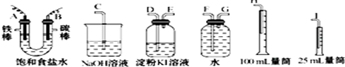
(2)设计上述气体实验装置时,各接口的正确连接顺序为:A接______、______接______;B接______、______接______.

(3)证明产物中有Cl2的实验现象是______.

(4)已知电解后测得产生的H2的体积为44.8mL(已经折算成标准状况),电解后溶液的体积为50mL,此时溶液中NaOH的物质的量浓度为:______.

答案

(1)铁是活泼金属,如果作阳极,在电流的作用下,铁失电子的能力大于氯离子失电子的能力,所以电解时不能得到氯气,故铁只能作阴极;碳棒是惰性电极,作阳极.

电解饱和食盐水时,氢离子得电子能力大于钠离子,所以在阴极即A极上氢离子得电子生成氢气,发生还原反应,电极反应式为2H++2e-=H2↑;

氯离子失电子能力大于氢氧根离子,所以在阳极上即B极上氯离子失电子生成氯气,发生氧化反应,电极反应式为:2Cl--2e-=Cl2↑;

故答案为:2H++2e-=H2↑;2Cl--2e-=Cl2↑.

(2)A极上产生的是氢气,用排水法收集氢气,因为氢气的密度小于水的,所以要采用向下排水法收集,即短导管为进气管,长导管为出水管,所以连接顺序为A→G→F;因为收集的氢气体积大于25mL,所以要用100mL的量筒收集水,所以F连接H;

B极上产生的气体是氯气,要检验氯气,可通过淀粉碘化钾溶液检验,氯气有强氧化性,能和碘化钾反应生成碘,碘遇淀粉变蓝色,氯气的密度小于碘化钾溶液的密度,所以长导管为进气管,短导管为出气管;氯气有毒,直接排空污染大气,且氯气和碱反应生成无毒物质,所以可用碱液吸收多余的氯气,所以连接顺序为B→D→E→C..

故答案为:A→G→F→H;B→D→E→C..

(3)因为Cl2+2KI=I2+2KCl,碘遇淀粉变蓝色,所以观察到的现象是淀粉碘化钾溶液变蓝色.

故答案为:淀粉碘化钾溶液变蓝色.

(4)2NaCl+2H2O=Cl2↑+H2↑+2NaOH 

                       22.4L     2mol        

                       0.0448L     0.004mol

C=

n
V
=
0.004mol
0.05L
=0.08mol/L

故答案为:0.08mol/L

单项选择题
单项选择题

Scientists studying the activity of the living brain with widely used new imaging techniques have been missing some of the earliest steps in brain activity because those changes are subtle and are masked by reactions that happen seconds later, Israeli scientists say.
The imaging techniques — positron emission tomography scanning and magnetic resonance imaging, known as PET and functional M. R. I. scans — are used prominently in studies of brain activity. The most active brain areas appear to light up on the scans as specific tasks are performed. The two techniques do not measure nerve-cell activity directly; they measure the extra flow of blood that surges to the most active brain areas.
Researchers at the Weizmann Institute of Science in Rehovot, Israel, have monitored these changes in blood flow in anesthetized cats by removing parts of the skull and observing how the nerve cells in activated regions fuel their activities by rapidly removing oxygen from nearby red blood cells.
This rapid uptake of oxygen, made evident by visible changes in the color of the red cells, proves that early oxygen transfer gives these neurons the energy to do their work, the researchers said.
They also found that subtle changes in blood flow began significantly earlier than was detected by PET and functional M. R. I. scans, which lack sufficient resolution and do not form their images quickly enough to follow such rapid changes. Dr. Amiram Grinvald published the findings in the Journal Science.
"The initial event is very localized and will be missed if you don’t look for it soon enough and use the highest possible resolution," Dr. Grinvald said. "Now people are beginning to use our results with other imaging methods."
Working on the exposed brain lets researchers follow electrical activity and the accompanying blood flow in greater detail than is possible by using indirect imaging methods that track neural activity through the skull. However, opportunities for open-skull studies of humans are limited to some kinds of neurosurgery, and researchers must mostly rely on PET and functional M. R. I. images for studies linking behavior with specific brain activity.
By directly observing exposed cat brains and in similar work with a few human cases, Dr. Grinvald and his associates have been able to observe the first evidence of electrical activity and other changes in brain cells after a light has been seen or a limb moved.
The newest research showed that it took three seconds or more after an event for the flow of blood to increase to an area of the brain dealing with a stimulus. That is the blood-flow increase usually pictured in brain-function studies with PET or functional M. R. I techniques, the Israeli researchers said. However, the initial reaction observed in the Weizmann research by directly imaging the exposed brain — the direct transfer of oxygen from blood cells to neurons — occurred in the first-tenth of a second and was lost to conventional imaging, they said.
The later increase in blood flow to the area, Dr. Grinvald said, was obviously an attempt by the body to supply more oxygen for brain activity. But the increase in blood was so abundant that it covered an area much larger than the region directly involved in the activity being studied, masking some of the subtle changes, he said.
The body’s reaction, the researchers said in the paper, was like "watering the entire garden for the sake of one thirsty flower."
Dr. Kamil Ugurbil, said that the Israeli research provided clues that allowed the use of functional M. R. I. scans to picture earlier events in the activity of brain cells.
"Dr. Grinvald’s observations are very important, and they have significant implications for functional imaging with high resolution," Dr. Ugurbil said in an interview. "We have actually been able to look at the early changes with magnetic resonance imaging, but you need to use higher magnetic fields to see them clearly because they are small effects."
By timing their images more carefully and by using per magnetic fields than normal, he said, researchers have used Dr. Grinvald’s findings to study early neuronal responses to stimuli at smaller, more specific sites in the brain.

According to the passage, scientists can’t observe some of the earliest steps in brain activity because ______ .

A.those changes are subtle and masked by some reactions

B.subtle changes in blood flow began earlier

C.the imaging techniques are out of place

D.the flow of blood to increase to an area of the brain is slow