问题 问答题

(一)

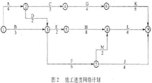
某综合楼工程,地下1层,地上10层,钢筋混凝土框架结构,建筑面积28500m2,某施工单位与建设单位签订了工程施工合同,合同工期约定为20个月。施工单位根据合同工期编制了该工程项目的施工进度计划,并且绘制出施工进度网络计划见图2(单位:月)。

在工程施工中发生了如下事件:

事件一:因建设单位修改设计,致使工作K停工2个月。

事件二:因建设单位供应的建筑材料未按时进场,致使工作H延期1个月。

事件三:因不可抗力原因致使工作F停工1个月。

事件四:因施工单位原因工程发生质量事故返工,致使工作M实际进度延迟1个月。

问 题

上述事件发生后,本工程网络计划的关键线路是否发生改变?如有改变,指出新的关键线路。

答案

参考答案:

上述事件发生后,本工程网络计划的关键线路没有发生改变。

单项选择题
单项选择题

Scientists studying the activity of the living brain with widely used new imaging techniques have been missing some of the earliest steps in brain activity because those changes are subtle and are masked by reactions that happen seconds later, Israeli scientists say.
The imaging techniques — positron emission tomography scanning and magnetic resonance imaging, known as PET and functional M. R. I. scans — are used prominently in studies of brain activity. The most active brain areas appear to light up on the scans as specific tasks are performed. The two techniques do not measure nerve-cell activity directly; they measure the extra flow of blood that surges to the most active brain areas.
Researchers at the Weizmann Institute of Science in Rehovot, Israel, have monitored these changes in blood flow in anesthetized cats by removing parts of the skull and observing how the nerve cells in activated regions fuel their activities by rapidly removing oxygen from nearby red blood cells.
This rapid uptake of oxygen, made evident by visible changes in the color of the red cells, proves that early oxygen transfer gives these neurons the energy to do their work, the researchers said.
They also found that subtle changes in blood flow began significantly earlier than was detected by PET and functional M. R. I. scans, which lack sufficient resolution and do not form their images quickly enough to follow such rapid changes. Dr. Amiram Grinvald published the findings in the Journal Science.
"The initial event is very localized and will be missed if you don’t look for it soon enough and use the highest possible resolution," Dr. Grinvald said. "Now people are beginning to use our results with other imaging methods."
Working on the exposed brain lets researchers follow electrical activity and the accompanying blood flow in greater detail than is possible by using indirect imaging methods that track neural activity through the skull. However, opportunities for open-skull studies of humans are limited to some kinds of neurosurgery, and researchers must mostly rely on PET and functional M. R. I. images for studies linking behavior with specific brain activity.
By directly observing exposed cat brains and in similar work with a few human cases, Dr. Grinvald and his associates have been able to observe the first evidence of electrical activity and other changes in brain cells after a light has been seen or a limb moved.
The newest research showed that it took three seconds or more after an event for the flow of blood to increase to an area of the brain dealing with a stimulus. That is the blood-flow increase usually pictured in brain-function studies with PET or functional M. R. I techniques, the Israeli researchers said. However, the initial reaction observed in the Weizmann research by directly imaging the exposed brain — the direct transfer of oxygen from blood cells to neurons — occurred in the first-tenth of a second and was lost to conventional imaging, they said.
The later increase in blood flow to the area, Dr. Grinvald said, was obviously an attempt by the body to supply more oxygen for brain activity. But the increase in blood was so abundant that it covered an area much larger than the region directly involved in the activity being studied, masking some of the subtle changes, he said.
The body’s reaction, the researchers said in the paper, was like "watering the entire garden for the sake of one thirsty flower."
Dr. Kamil Ugurbil, said that the Israeli research provided clues that allowed the use of functional M. R. I. scans to picture earlier events in the activity of brain cells.
"Dr. Grinvald’s observations are very important, and they have significant implications for functional imaging with high resolution," Dr. Ugurbil said in an interview. "We have actually been able to look at the early changes with magnetic resonance imaging, but you need to use higher magnetic fields to see them clearly because they are small effects."
By timing their images more carefully and by using per magnetic fields than normal, he said, researchers have used Dr. Grinvald’s findings to study early neuronal responses to stimuli at smaller, more specific sites in the brain.

According to the passage, scientists can’t observe some of the earliest steps in brain activity because ______ .

A.those changes are subtle and masked by some reactions

B.subtle changes in blood flow began earlier

C.the imaging techniques are out of place

D.the flow of blood to increase to an area of the brain is slow